Why Laser Treatment for Melasma and Sunspots Depends on Whether Pigment Sits in the Epidermis or Dermis
By Dr. Lee7 min read
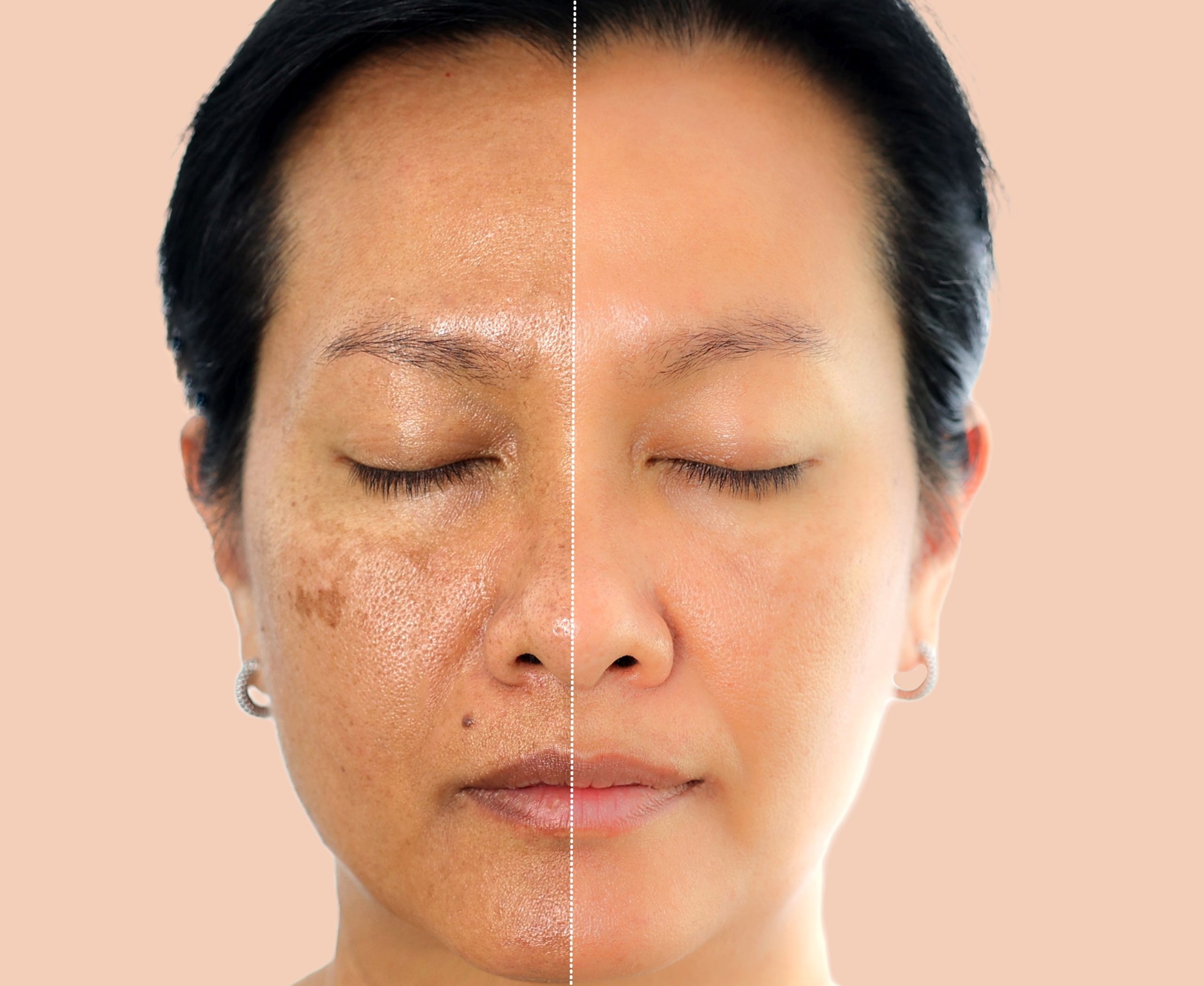
You catch a brown patch in the mirror, melasma or a sunspot, something you have probably noticed before. But two spots that look the same shade of brown can be sitting in completely different places under the skin. Some pigment lives just beneath the surface. Other pigment sits much deeper. And that single difference in depth ends up being the biggest factor in why one treatment works well and another barely makes a dent. This is why, when a clinic evaluates pigment, the color and size are only part of the picture. What matters just as much is whether that pigment leans toward the epidermis or the dermis. Two spots can look nearly identical on the surface, but if what is happening underneath is different, the approach needs to be different too.
Where Does Pigment Actually Form, the Epidermis or the Dermis?
Skin is built from two main layers. The outer layer is the epidermis, and beneath it sits the dermis. Think of the epidermis as wallpaper and the dermis as the wall structure behind it. If a stain gets on the wallpaper, wiping the surface or swapping the paper solves it. But if a stain has soaked into the plumbing behind the wall, changing the wallpaper alone does nothing.
Pigment works the same way. Heavy sun exposure or repeated inflammation pushes melanocytes, the cells that produce your skin color, to churn out excess melanin. When that pigment stays confined to the epidermis, it is epidermal pigmentation. When it gradually seeps down into the dermis over time, it becomes dermal pigmentation. Melasma, age spots, freckles, and post-inflammatory pigmentation can all look similar from the outside, but what actually differs between them is which of these two layers holds more of the pigment.
Why Does Epidermal Pigment Respond So Quickly to Treatment?
The epidermis is the shallowest layer of skin, so it is thin. Pigment sitting inside it stays close to the surface, and the cycle of cells being made and shed, what dermatologists call turnover, moves fast in this layer. Epidermal cells typically replace themselves roughly every 4 weeks, so simply reducing whatever is triggering pigment production can sometimes let a spot fade on its own.
Treatment has the advantage here too. Brightening ingredients like hydroquinone and tranexamic acid work by suppressing the process melanocytes use to make pigment in the first place, and because the epidermis is so close to the surface, these ingredients do not have far to travel to take effect. The same logic applies to lasers. Pigment sitting in the epidermis can be broken down even when the laser wavelength only reaches a shallow depth, so relatively gentle settings tend to produce good results.
Why Is Dermal Pigment So Much Harder to Treat?
The dermis is far thicker than the epidermis and packed densely with blood vessels, nerves, and collagen. Once pigment migrates this deep, the situation changes entirely. Pigment found in the dermis is usually not something melanocytes produced directly there. More often, it is melanin that leaked down from the epidermis and got swallowed up by macrophages, the immune system's cleanup cells, which then hold onto it for a long time.
The problem is that these macrophages do not turn over quickly the way epidermal cells do. Once they settle in, they can sit there unchanged for months or even years. As a result, dermal pigment fades on its own far more slowly than epidermal pigment, and topical brightening ingredients struggle to reach the dermis in a strong enough concentration to make much difference. It is the same idea as only cleaning the surface of a wall while the stain has already soaked into the pipes behind it.
What Treatment Principles Work for Epidermal Pigment?
Epidermal pigmentation responds best to methods that act near the surface. Topical brighteners, exfoliating peels, and low-fluence toning lasers are the go-to options.
- Topical brighteners: Ingredients like hydroquinone, tranexamic acid, and niacinamide dial down the signal melanocytes use to produce pigment. Because they only need to work within the epidermis, a short penetration distance is enough to get meaningful results.
- Peels: These thin out the stratum corneum and force epidermal turnover to speed up. The idea is to help old, pigment-carrying skin cells shed off faster than they normally would.
- Low-fluence toning lasers: These break down epidermal melanin gradually using low energy delivered across multiple sessions. Rather than burning through the pigment in one aggressive pass, this spread-out approach suits pigment confined to the epidermis.
What Treatment Principles Work for Dermal Pigment?
Because surface treatments barely reach dermal pigment, deeper-penetrating wavelengths or lasers that target pigment directly are needed instead. Q-switched lasers and picosecond lasers are the most common examples. These deliver intense energy to pigment particles in an extremely short burst, shattering the pigment into tiny fragments without spreading heat damage into the surrounding tissue.
Once pigment is fragmented this way, the body's immune cells clear it out gradually. Macrophages swallow up the broken pigment particles and carry them off through the lymphatic system, which is why results from dermal pigment treatment often take longer to become visible compared to epidermal pigment. The dermis is also rich in blood vessels and nerves, making it more sensitive to irritation, and an ill-suited setting can actually trigger new inflammatory pigmentation within the dermis. That is why choosing the right wavelength and power requires extra care in this layer.
How Can Depth Be Assessed Before Treatment?
Even with the same brown spot, telling epidermal from dermal, or a mixed type where both layers are involved, is hard to do with the naked eye alone. In practice, clinics commonly rely on a Wood's lamp, a specialized ultraviolet light examination, or a dermatoscope to gauge depth by examining a spot's borders and tone.
- Epidermal type: The border tends to look fairly well defined, and the pigment appears darker and more pronounced under a Wood's lamp. This happens because pigment concentrated in a shallow layer creates a bigger contrast in how light reflects back.
- Dermal type: The border tends to look blurred, and there is little visible change under a Wood's lamp. Since the pigment sits deeper, light does not reflect back strongly enough to show much contrast.
- Mixed type: Both characteristics show up together, and in actual clinical practice, this mixed pattern is quite common.
What's Easy to Overlook When Planning Treatment?
One common misconception in pigment treatment is starting dermal pigment off with an aggressive epidermal-style approach and only switching methods if it fails to work. But the dermis is a sensitive layer, and repeated irritation from a mismatched method can trigger new pigmentation instead of clearing the existing spot. That is why choosing the depth-appropriate method from the very beginning tends to pay off far more in the long run.
Another thing worth setting expectations around is speed. Epidermal pigment can sometimes show visible change within a few weeks, but dermal pigment naturally takes longer to metabolize, so it often fades gradually across multiple sessions. If progress feels slow midway through, understanding that dermal pigment simply moves at its own pace makes the process much easier to sit through.
Sun protection matters equally for both epidermal and dermal pigmentation. UV exposure is the single biggest trigger for re-stimulating melanocytes into producing new pigment, so keeping treated spots from returning depends on consistent follow-up care. Reapplying sunscreen every day will not erase pigment that has already formed, but it does reduce the signal telling melanocytes to make more. Because of this, treatment and sun protection are best thought of not as separate steps but as one continuous process, and results tend to hold up far longer when they are managed together.
It is also worth noting that pigment depth can differ across different areas of the same face. Areas like the cheekbones that get prolonged sun exposure often have pigment that has migrated deep into the dermis, while less exposed areas tend to keep pigment confined to the epidermis. Because of this, different settings may be needed for different areas of the same face, and figuring out that plan always starts with checking the pigment's depth first.
References
Korean Dermatological Association, American Academy of Dermatology (AAD), and Ministry of Food and Drug Safety (MFDS).
Was this helpful?
About this article
Written by a practising aesthetic physician and intended for general education — not a substitute for individual medical advice.
Read next

Freckles and Melasma Look Alike, So Why Do Doctors Treat Them in Opposite Ways?
At a glance, freckles and melasma both look like brown spots, which makes them easy to mix up. But where the pigment actually sits in the skin, and why it shows up in the first place, are completely different. That difference decides whether you need a strong laser or a gentle toning session, and why one of the two keeps coming back.
By Dr. Lee
Can Lasers Fade a Cafe-au-Lait Spot? Telling It Apart From Moles and Melasma, and Why It Keeps Coming Back
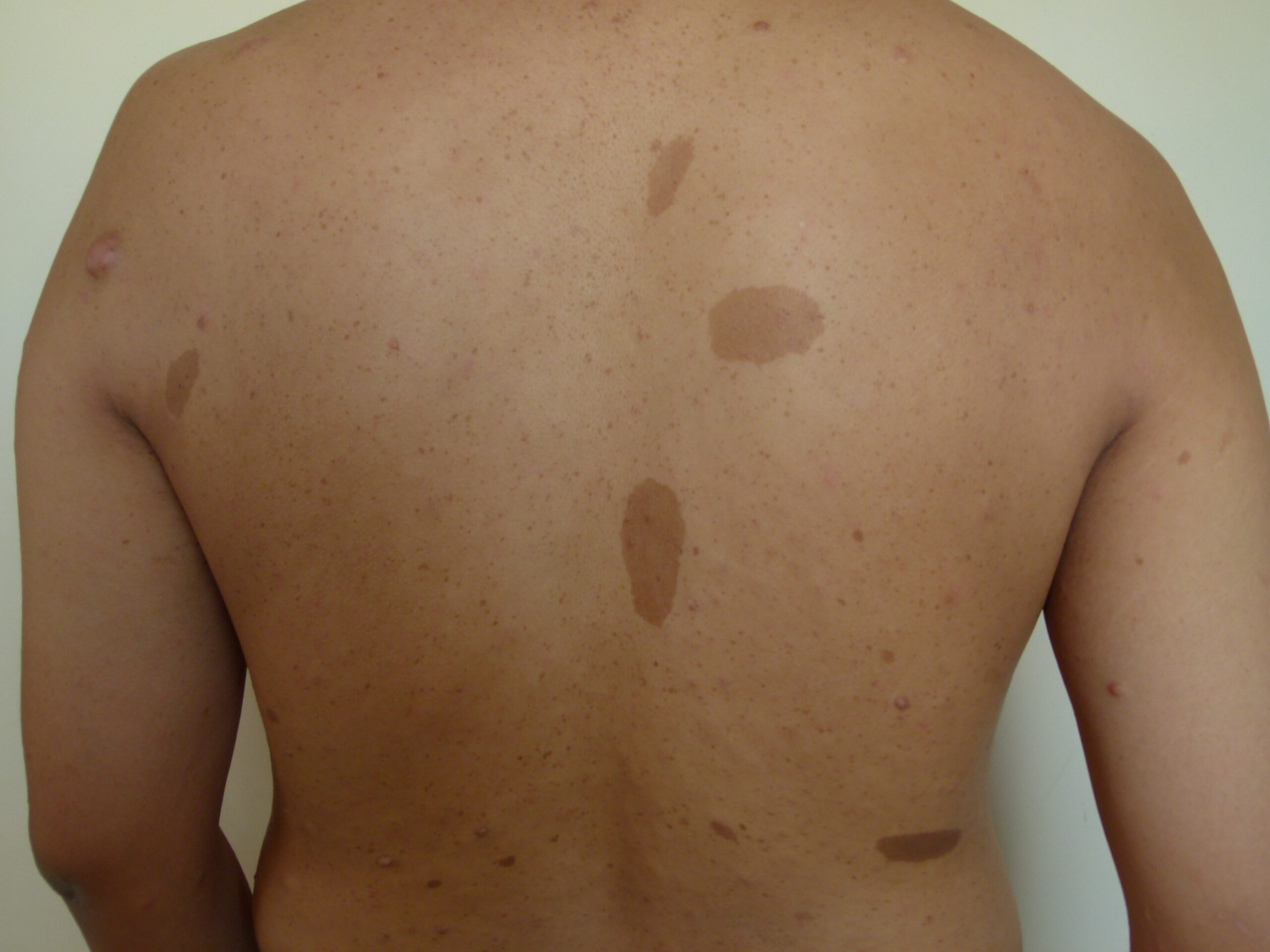
A cafe-au-lait spot is a light milk-coffee brown patch on the cheek or arm, and because its pigment sits in the shallow epidermis, laser light reaches it easily, though it also tends to come back. Here's how to tell it apart from moles and melasma, why multiple spots call for a check-up, how much laser treatment can fade it, and how to manage recurrence.
By Dr. Kim

ABNOM: Why Creams Can't Clear It and What the Laser Data Show
ABNOM (acquired bilateral nevus of Ota-like macules) sits in the dermis, beyond the reach of brightening creams or oral supplements. Here's what separates it from melasma, why Q-switched laser is the standard treatment, and what clinical data say about session count, clearance rates, and the real risk of post-treatment darkening.
By Dr. Lee