Nevus of Ota: How Many 1064 nm Laser Sessions It Takes to Clear Blue-Grey Dermal Pigment, and the Risk of Recurrence
By Dr. Kim8 min read

If a dark, bluish patch has spread across one eye area or cheekbone, it may not be melasma or ordinary discoloration at all. That's especially true if it's been there since birth or deepened around puberty. Nevus of Ota carries its pigment deep in the dermis rather than at the skin's surface, which is exactly why topical brightening creams rarely make a visible dent.
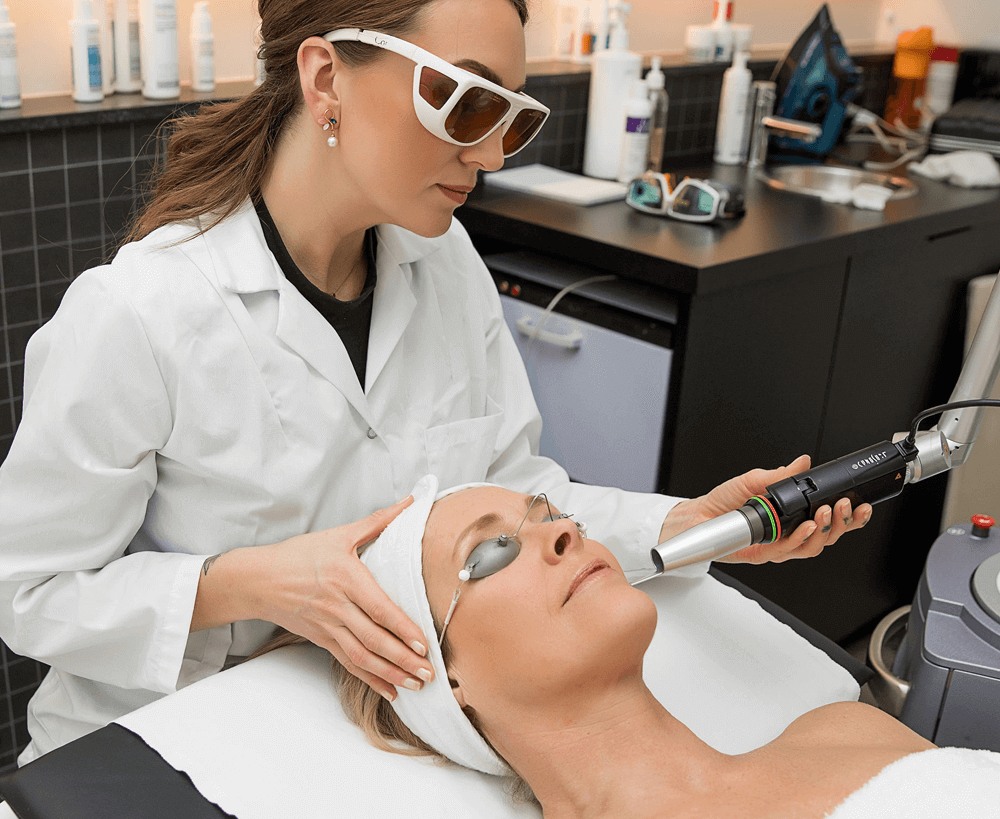
That's why the standard treatment for nevus of Ota isn't a cream or an oral medication, it's laser. A 1064 nm Q-switched Nd:YAG laser reaches the deep dermal pigment, breaks the clumps into smaller pieces, and lets the body clear them out gradually over multiple sessions. You're probably wondering how much improvement to expect after a given number of sessions, and whether recurrence or pigment side effects are a real concern. Below, we'll also cover how to tell it apart from the similar-looking acquired bilateral nevus of Ota-like macules (ABNOM).
What Kind of Pigment Disorder Is Nevus of Ota?
Nevus of Ota is a dermal melanocytosis, meaning the pigment-producing cells sit in the dermis instead of the epidermis. During development, melanocytes that should have migrated up to the epidermis get left behind in the dermis instead. Because that pigment sits deeper, light passing through it scatters differently, so instead of reading as brown, it appears bluish or slate-grey.
The distribution follows a distinct pattern. It runs along the first and second branches of the trigeminal nerve, the nerve responsible for facial sensation. That means it shows up around the eyes, forehead, cheekbones, and bridge of the nose, almost always on just one side of the face. It's more common in women, with a reported ratio of roughly 5 to 1, and while it's usually present from birth, it can also become more noticeable during hormonal shifts like puberty or pregnancy.
It can affect the eye as well. In about two-thirds of patients, pigment also appears on the sclera, the white of the eye. That area is difficult to treat with laser, so it's safer to have an ophthalmologist monitor it as well. On the surface, nevus of Ota is easy to mistake for melasma or ordinary spots, but because the depth and distribution of the pigment are entirely different, the treatment approach differs too. So the first step, before any treatment begins, is pinning down exactly how deep the pigment sits.
How Is Nevus of Ota Different from ABNOM?
The condition most often confused with nevus of Ota is acquired bilateral nevus of Ota-like macules, known by the acronym ABNOM. Both involve dermal pigment that reads bluish and both respond to 1064 nm laser, but they differ in when they appear and how they're distributed.
| Feature | Nevus of Ota | ABNOM |
|---|---|---|
| Onset | Congenital or puberty | Acquired, adulthood |
| Laterality | Unilateral | Bilateral, symmetric |
| Main sites | Eye area, forehead, cheekbones, nose bridge | Both cheekbones |
| Scleral involvement | Possible | Absent |
| Pigment depth | Dermal | Dermal |
The biggest difference is symmetry. Nevus of Ota usually shows up on just one side of the face, while ABNOM appears symmetrically on both cheekbones in adulthood. Nevus of Ota also frequently involves pigment on the white of the eye, while ABNOM never touches the sclera or oral mucosa.
Timing helps distinguish them too. Nevus of Ota is present from birth or deepens during puberty, while ABNOM almost always first develops sometime in your twenties or later. The two conditions can occasionally coexist on the same face, so it's worth looking at both the timing and the distribution of the pigment together before mapping out a treatment plan. Rather than jumping straight into aggressive laser sessions based on a guess, it's safer to first pin down exactly which condition you're dealing with.
Why Does It Take Multiple Rounds of 1064 nm Laser?
The pigment in nevus of Ota sits deep in the dermis. Topical brighteners and oral medications simply can't reach that depth, which is why they have so little effect. Clearing dermal pigment requires something that can reach that depth and physically break the pigment apart, and that's the role laser plays.
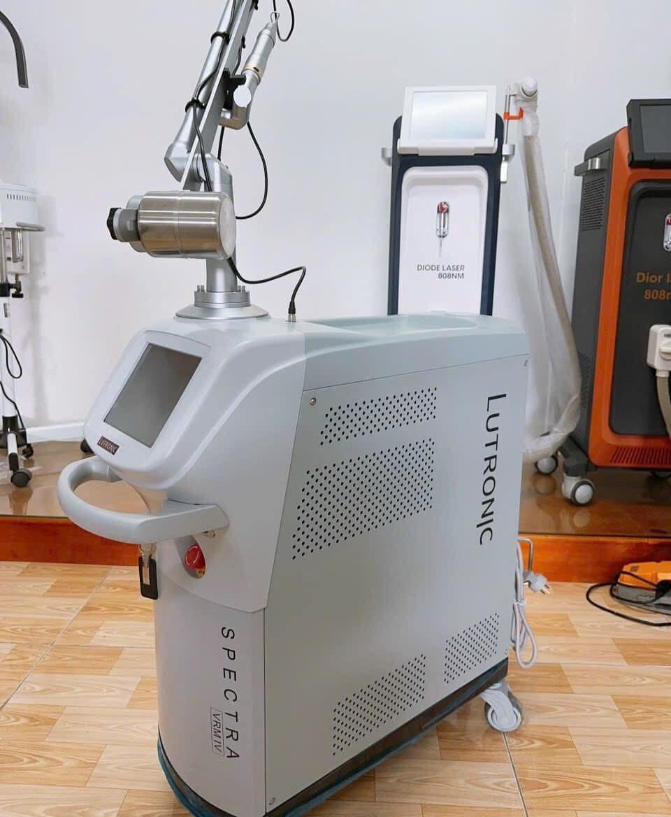
That's also why 1064 nm is the wavelength of choice here. Longer wavelengths penetrate deeper into skin, so 1064 nm light passes through the epidermis and reaches the melanocytes sitting in the dermis. At the same time, it's absorbed less by epidermal melanin, so it irritates the skin's surface less while selectively targeting the deeper pigment. The Q-switched delivery fires an intense burst of light in an extremely short pulse, breaking up pigment clumps with a force closer to a pressure wave than heat.
There's also a reason a single session isn't enough. The shattered pigment gets slowly consumed by immune cells in the skin and carried away through the lymphatic system, and that cleanup process takes time. On top of that, the melanocytes in the dermis keep producing pigment between treatments, so it takes several sessions spaced weeks to months apart for the color to fade in stages. Unlike surface spots that slough off with dead skin cells, it helps to think of dermal pigment treatment as a repeated process of gradually chipping away at the color.
How Many Sessions Does It Take, and How Much Improvement Can You Expect?
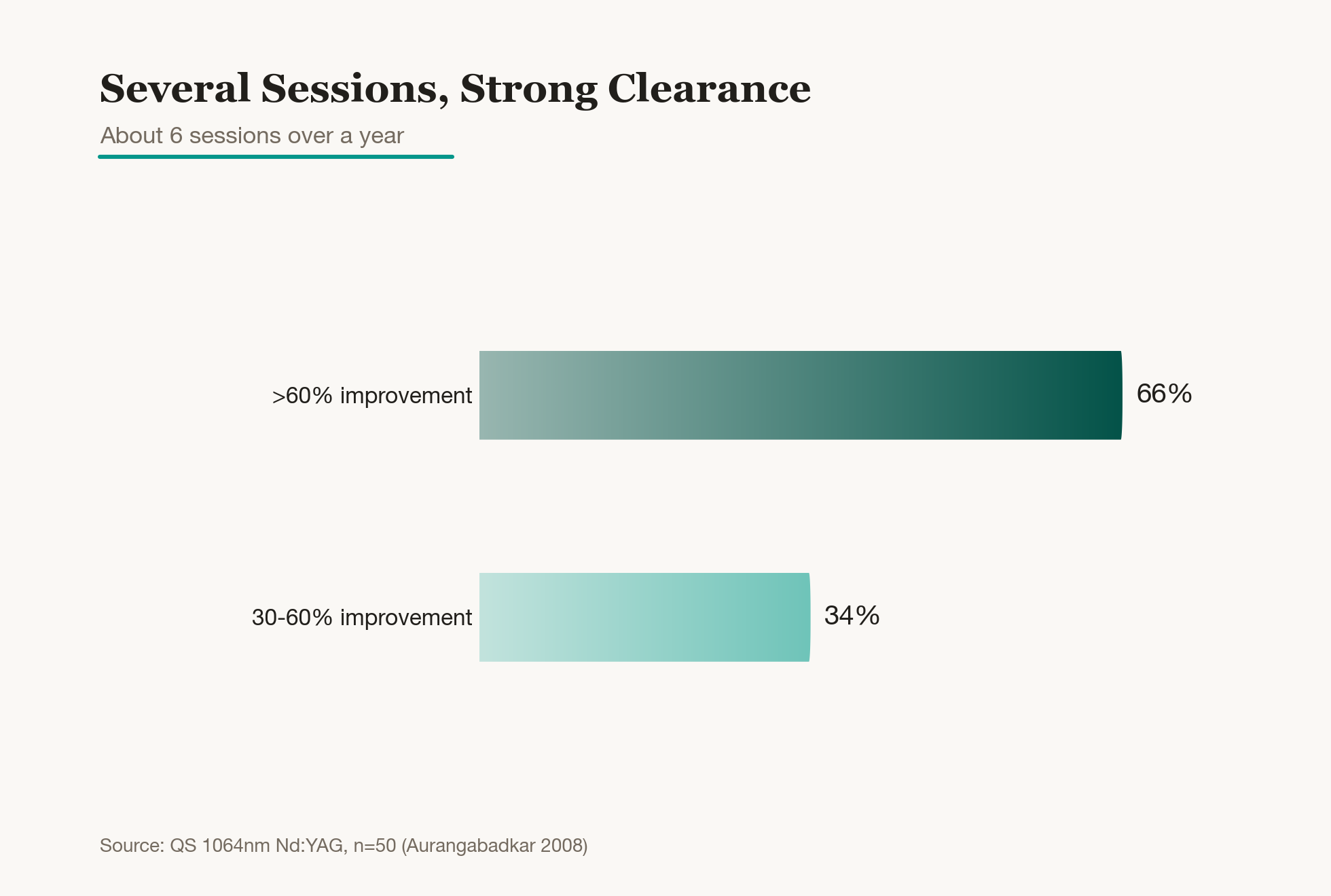
The relationship between session count and outcome has been studied fairly well. In a study that treated 50 patients with nevus of Ota using a 1064 nm Q-switched Nd:YAG laser, patients averaged 6 sessions over a year, and 66% saw more than 60% improvement. The remaining patients still saw improvement in the 30% to 60% range, with no non-responders at all.
The key takeaway is that this treatment requires patience. Dermal pigment is deep and stubborn, so it doesn't disappear after one or two sessions, it fades slowly across many. Multiple studies have found that more sessions correlate with better results, and that starting treatment at a younger age tends to produce a better response. So rather than leaving it untreated for years, it's usually better to get an accurate diagnosis and start treatment early.
It helps to set expectations realistically from the start. Because the fading happens gradually, session by session, it helps to go in knowing this is a multi-session process, rather than giving up early because the change after one or two rounds seems small. Keep at it consistently, spaced weeks to months apart, and the pigment becomes noticeably lighter over time. Satisfaction tends to run high once patients complete the full course, so it's worth planning for the long haul from the outset.
Can Pigment Get Worse, or Come Back After Treatment?
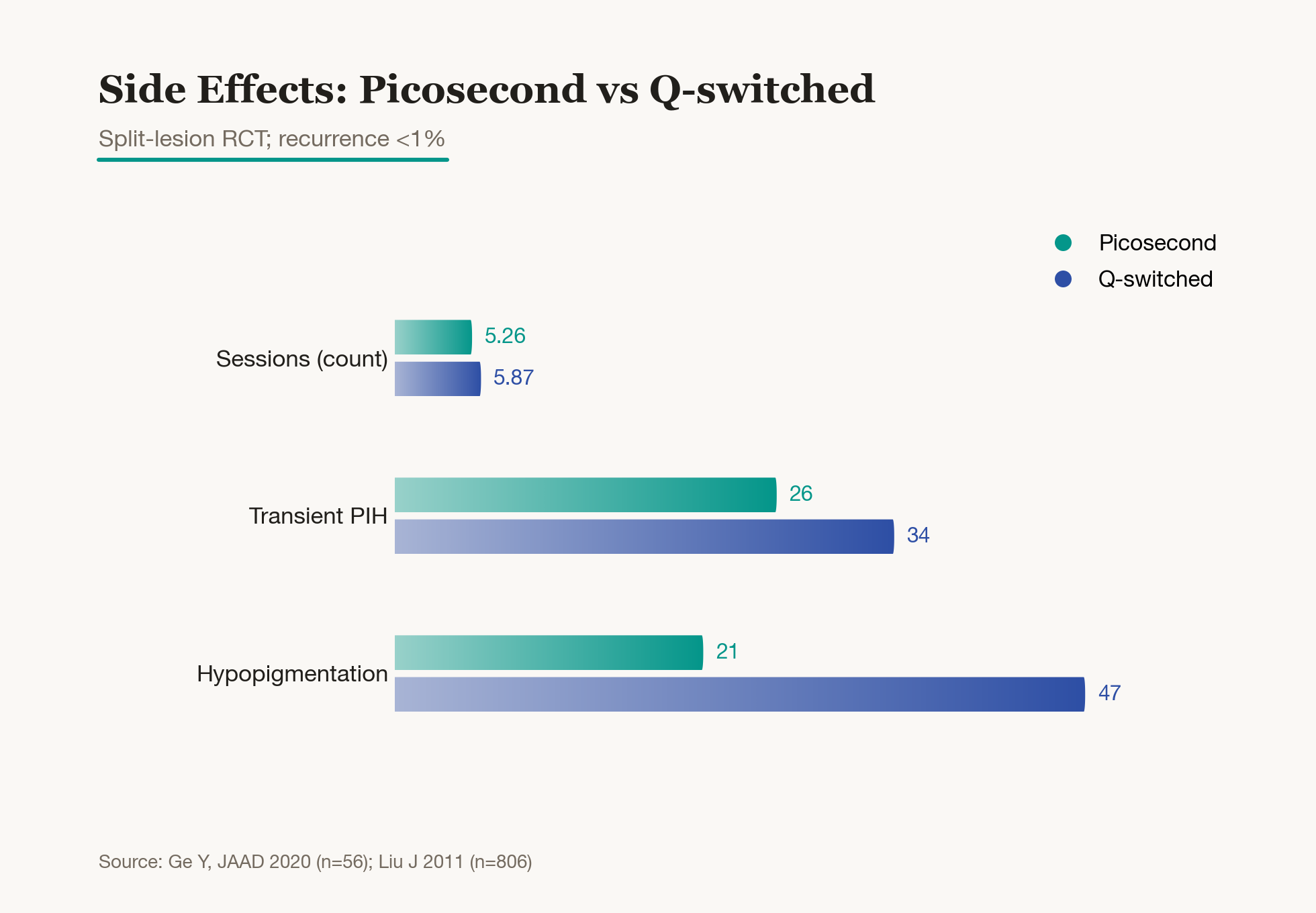
Breaking down deep pigment with laser can come with a temporary side effect: pigment that looks darker for a while, known as post-inflammatory hyperpigmentation. It's usually short-lived, though. In the 50-patient study mentioned above, 10% developed this kind of darkening, but it faded within two months with sun protection and brightening care. The opposite can also happen, temporary lightening called hypopigmentation, so using conservative energy settings and properly spacing out sessions is the safer approach.
Picosecond lasers have recently widened the options. In a study that split nevus of Ota patches in half and compared Q-switched laser against picosecond laser, the picosecond side needed slightly fewer sessions on average and produced fewer side effects like hyper- and hypopigmentation. The principle is that shattering pigment in an even shorter pulse reduces surrounding irritation. That said, both approaches are effective, standard treatments for nevus of Ota, so choosing between them comes down to what fits your situation.
Recurrence is uncommon. In a long-term study that followed over 800 patients, recurrence after treatment was under 1%. Still, there are rare reports of pigment gradually returning over time, so it's worth continuing consistent sun protection even after treatment ends, and returning for a follow-up if you notice any changes.
So What's the Best Way to Approach This?
Nevus of Ota may resemble melasma or ordinary spots on the surface, but it's an entirely different pigment disorder with its color rooted deep in the dermis. Topical or oral treatments have real limits here, which is why multiple sessions of 1064 nm Q-switched Nd:YAG laser form the core of treatment. Picosecond laser can be considered alongside it depending on the case.
| Item | Key point |
|---|---|
| Standard treatment | 1064 nm Q-switched laser |
| Sessions | Multiple, spaced out |
| Picosecond laser | Somewhat fewer sessions, fewer side effects |
| Recurrence | Uncommon |
| Aftercare | Sun protection |
Before starting, the first priority is confirming whether it's melasma or nevus of Ota, or whether ABNOM might be involved as well. Once treatment begins, because dermal pigment is deep and stubborn, you'll need patience to see the gradual, multi-session fading process through. Pigment can temporarily darken along the way, so sticking to a safe interval and energy setting, rather than pushing intensity, ends up being the faster path.
Aftercare matters just as much once treatment wraps up. UV exposure is the biggest trigger for re-stimulating pigment, so thorough sun protection during and after treatment is well worth the effort. Nevus of Ota delivers its most satisfying results when accurate diagnosis, patiently pursued multi-session laser treatment, and diligent aftercare all come together. Knowing from the start that this is a treatment that takes time makes it much easier to stay the course without losing patience halfway through.
Was this helpful?
About this article
Written by a practising aesthetic physician and intended for general education — not a substitute for individual medical advice.
Read next

ABNOM: Why Creams Can't Clear It and What the Laser Data Show
ABNOM (acquired bilateral nevus of Ota-like macules) sits in the dermis, beyond the reach of brightening creams or oral supplements. Here's what separates it from melasma, why Q-switched laser is the standard treatment, and what clinical data say about session count, clearance rates, and the real risk of post-treatment darkening.
By Dr. Lee

Why Does Laser Hair Removal Require Multiple Sessions? Session Guidelines Based on Growth Cycles and the Alexandrite Wavelength
Laser hair removal doesn't wrap up in one or two visits, and here's why: it comes down to the growth cycle that alternates between an active growth phase and a resting phase. We'll walk through how many sessions each area typically needs and how the alexandrite and Nd:YAG wavelengths differ.
By Dr. Kim
How Q-switched Laser Toning Clears Pigment With Repeated Low-Energy Pulses, and the Hypopigmentation Risk to Watch
Laser toning fires low energy again and again instead of one strong blast, and there is a real reason for that. Here is the pigment-breakdown science behind it, how overtreatment can trigger side effects like hypopigmentation, and how to keep session spacing safe.
By Dr. Lee