CO2 Pinhole Laser for Under-Eye Syringoma: How Many Sessions Until It Fades?
By Dr. Kim10 min read

The small, grainy bumps under the eyes known as syringoma, and the yellowish patches that show up on the eyelids called xanthelasma, are notoriously hard to cover with makeup. One option for treating these raised lesions is the CO2 pinhole method. Pinhole means exactly what it sounds like: a hole the size of a pin. The technique uses a laser to burn a dense grid of very narrow, deep holes straight into the lesion, destroying the tissue on the spot.
Unlike treatments that resurface an entire area at once, the pinhole method only punctures the lesion itself, point by point. The normal skin between the holes stays intact, which means faster healing and less scarring risk. That's why it's often considered for areas like the face, where scarring is something you really want to avoid. This article walks through how the pinhole method actually works, what kind of clearance you can realistically expect for syringoma and telangiectasia, and what recovery and pigmentation risk look like, based on the evidence.
How does the pinhole method remove a lesion?
A CO2 laser emits light at a 10,600nm wavelength, which happens to be absorbed extremely well by water. Since skin is mostly water, the moment this light hits it, the tissue at that spot vaporizes almost instantly. This tissue-vaporizing effect is called ablation.
The pinhole method doesn't spread this laser across a wide surface. Instead, it burns a dense grid of narrow, deep vertical holes spaced about 1 to 3mm apart directly over the lesion, similar to poking a row of holes into thick paper with an awl. Rather than just searing the surface, each hole drives down like a column into the lesion to break down the target tissue from within.
Here's how each hole is made. A single short laser pulse vaporizes the tissue at that exact spot, carving out one narrow vertical channel. The laser is moved slightly and fired again, repeating across the lesion to create a grid. Depth is adjusted based on how thick the lesion is, and what sets the pinhole method apart is that it doesn't just graze the surface. It reaches down to the mid-dermis. Raised, prominent lesions get deeper holes, and shallow ones get shallower holes.
These micro-holes do two things at once. First, they directly destroy the lesion tissue in their path. Second, the heat that spreads gently around each hole realigns collagen and elastin. Think of collagen as the pillars holding up the skin, and elastin as the rubber bands that pull it back into shape. As the two get rewoven, the skin tightens and the surface texture smooths out.
The key is that the normal skin between the holes stays completely intact. That surviving skin becomes the scaffold the wound heals against, since the surrounding cells migrate in from both sides to fill each gap. That's why the pinhole method causes less bleeding, heals faster, and carries a lower scarring risk than resurfacing an entire area at once.
The depth and spacing of the holes are adjusted to the lesion. A shallow, widespread lesion gets a dense grid of shallow holes, while a deep, firm one gets holes drilled a bit deeper. Being able to fine-tune the approach to the specific lesion this way is one of the real advantages of the pinhole method.
What other lesions is it used on besides syringoma?
| Lesion | Evidence for pinhole |
|---|---|
| Syringoma (under-eye bumps) | Solid evidence |
| Xanthelasma (yellow eyelid plaques) | Case reports |
| Telangiectasia | Studies available |
| Hypertrophic and burn scars | Studies available |
The pinhole method works best on lesions that sit raised above the surface or cluster in one spot. The classic example is syringoma, those small bumps that appear under the eyes. These are tiny growths from overactive sweat glands, and puncturing them directly removes the tissue. Xanthelasma, the yellow plaques that form on the eyelids from cholesterol buildup, is another lesion this technique is frequently tried on.
It's also used for telangiectasia, where fine red blood vessels show through the skin, and for hypertrophic scars that rise up after surgery or burns. In that case, the holes rework the collagen and press the raised scar down into something flatter. Broadly speaking, any lesion that stands out above the surface is worth considering for this approach.
One thing worth being upfront about: there's no dedicated study looking at the pinhole method specifically for enlarged pores. That said, since the heat around each hole reworks collagen and could theoretically tighten the pore walls, pores might end up looking firmer as a side effect. That's a mechanistic expectation at best, not something backed by a percentage improvement anyone can promise.
How much does syringoma fade after two sessions?
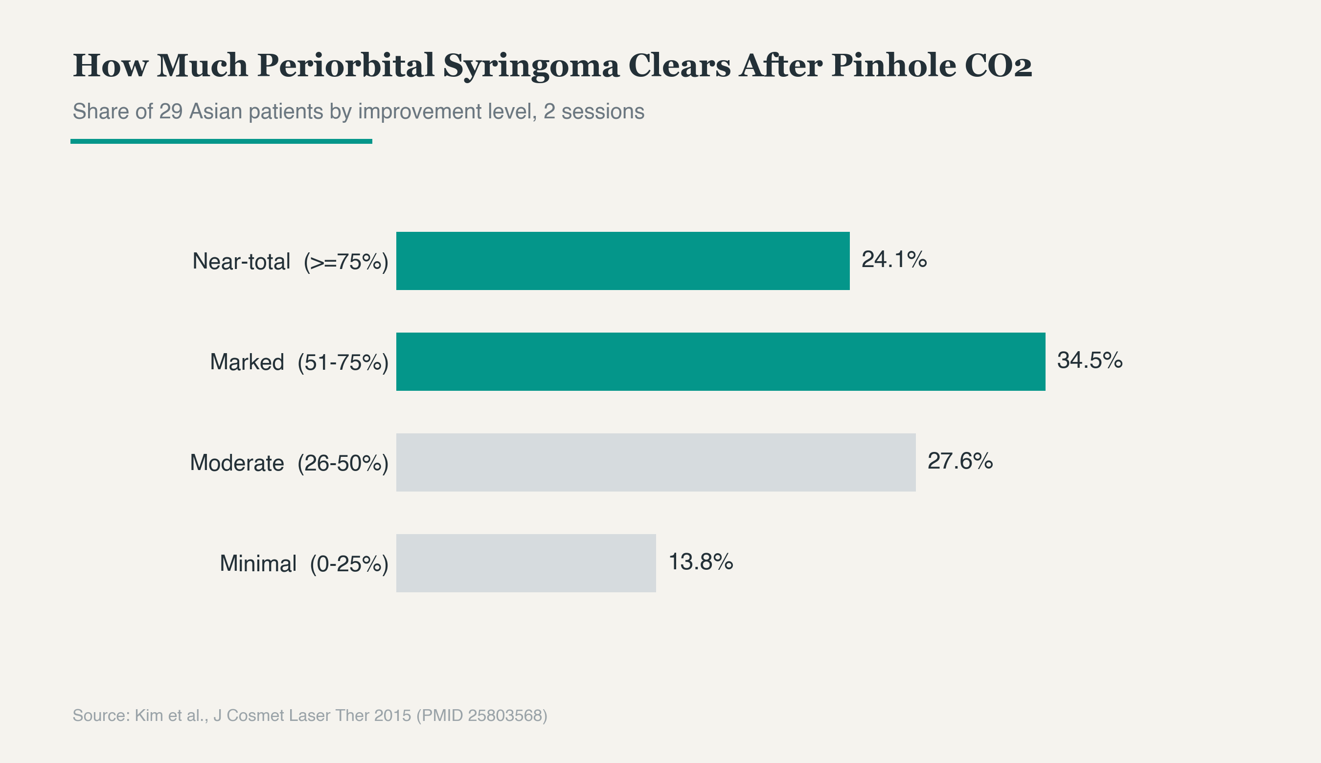
How much does syringoma actually improve with the pinhole method? After about two sessions on under-eye bumps, close to six out of ten patients saw more than half their lesions clear. As the graph above shows, some people saw near-total clearance, while others only saw a modest fade. Response varies from person to person, but overall, the raised bumps noticeably flatten out. And with each additional session, the improvement continues to build.
The numbers look solid on paper, but it's worth keeping expectations realistic. The pinhole method tends to fade lesions in stages rather than wiping them out in one go. Some people see a dramatic drop after a single session, while others need several rounds spread out over time before they're satisfied.
There's also the fact that syringoma ducts extend fairly deep into the skin, similar to a weed with roots that reach far underground. Clearing what's visible on the surface doesn't necessarily remove the entire duct, so recurrence over time is a real possibility. Recurrence is a genuine limitation here. It helps to go in expecting multiple sessions from the start and to think of the goal as making the bumps less noticeable rather than erasing them completely in one shot. That mindset alone makes the whole process feel much easier.
How is it different from fractional CO2?
| Pinhole method | Fractional CO2 | |
|---|---|---|
| Goal | Direct lesion removal | Broad resurfacing |
| Holes | Few, deep, on the lesion | Dense, shallow, across the area |
| Best for | Raised lesions like syringoma, scars | Photoaging, texture |
The names sound similar enough to cause confusion, but the two treatments are used quite differently. The pinhole method targets individual lesions directly. It uses fewer holes, but drills them deep, with the goal of removing raised tissue at its root. That makes it well suited to clearly defined, raised lesions like syringoma or scars.
Fractional CO2, by contrast, scatters a dense grid of shallow micro-holes across a broad area of skin. The goal isn't to eliminate a specific lesion, but to resurface and rejuvenate the skin over a wide area. That's why it's mainly used for dull, sun-damaged skin, fine texture refinement, or smoothing out subtle scar surfaces. Think of it as watering an entire lawn evenly to revive the whole thing.
The pinhole method works differently. Rather than spreading out, it digs deep into one spot to break down the lesion, closer to pulling a single weed out by its roots in a flower bed. In actual practice, clinicians often choose between the two based on the situation, or combine them. If a lesion is clearly raised, think pinhole. If the goal is refining the texture and tone of the whole surface, think fractional.
Scab period and pigmentation care
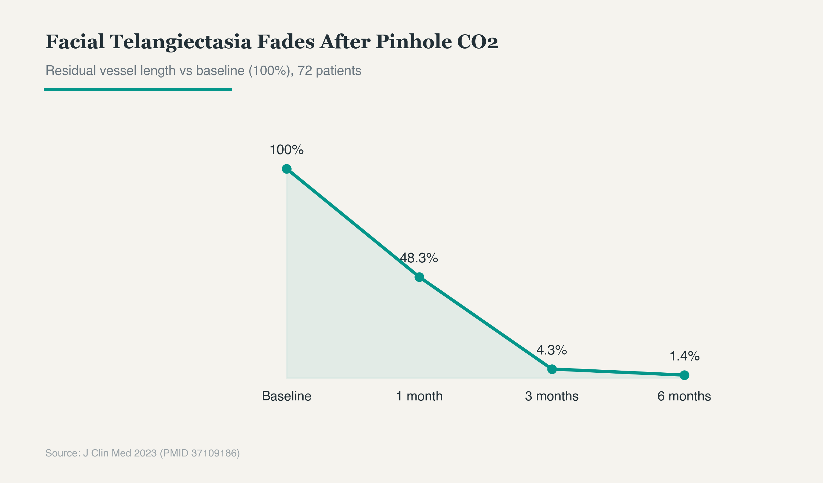
Recovery improves steadily over time. Take red blood vessels as an example: as the graph above shows, if you set the pre-treatment level at 100, it drops below half within a month and fades to almost invisible by around six months. The change might feel underwhelming at first, but it continues to resolve gradually over several months.
Downtime isn't especially heavy. Tiny pinpoint crusts form over the treated spots and last about 5 to 7 days, with redness lingering for another 1 to 2 weeks. If you get through this period carefully, the skin heals naturally on its own. It's best to let the crusts fall off on their own rather than picking at them, since forcing them off can leave marks or deepen pigmentation. During this stretch, it helps to keep the skin well moisturized and avoid anything irritating.
The thing that tends to cause the most concern is post-inflammatory hyperpigmentation, or PIH. This happens when the skin overproduces melanin as a defensive response while healing, leaving a darker patch behind. In this same study, 5 patients developed temporary pigmentation, but it resolved in all of them within six months. That said, Asian skin, which typically falls into Fitzpatrick types III to IV, tends to hold onto pigmentation more easily after an injury, which is why PIH is considered the main side effect to watch for. Even so, it's mostly a temporary reaction that fades with time. Keeping energy settings conservative and staying diligent with sun protection can meaningfully lower this risk. That's why it's worth applying sunscreen carefully whenever you head outside during recovery.
What to know before treatment
| To check | Details |
|---|---|
| Sessions | 2 for syringoma, 5-8 for scars |
| Interval | 2-3 months |
| Downtime | Crusting for 5-7 days |
| Watch for | Sun protection, possible recurrence |
A few things are worth confirming before deciding on treatment, since they make the whole experience feel a lot more manageable. First, it helps to understand how sessions work. Syringoma usually takes around 2 sessions, while scars can need anywhere from 5 to 8, spaced 2 to 3 months apart. Thinking of this as a gradual process handled in stages, rather than a single fix, makes it much easier to stay patient.
Sun protection matters a lot here too. Heavy sun exposure before or after treatment raises the risk of pigmentation, so applying sunscreen consistently afterward really matters. It's also worth keeping expectations realistic. Syringoma and xanthelasma are the kind of lesions that can come back even after they've improved, so it's more useful to think of this as fading them rather than eliminating them for good.
If your skin tone runs on the darker side, it's safer to try a small test patch before treating a larger area. Checking the response ahead of time helps lower the risk of pigmentation. When treating an area close to the eye, like the eyelid, protective eye shields are a must. These are small lens-like guards placed directly over the eye to block laser light from reaching it.
One last point: because this treatment targets lesions directly, the practitioner's hand skill plays a huge role in the outcome. How precisely they control the depth and spacing of each hole determines how recovery and results turn out. That's exactly why choosing an experienced practitioner matters for both the final result and your safety.
Was this helpful?
About this article
Written by a practising aesthetic physician and intended for general education — not a substitute for individual medical advice.
Read next
Reepot Laser Results: Can One or Two Sessions Clear Lentigines That Toning Couldn't?
How Reepot's 532nm wavelength achieves roughly 11x higher melanin absorption than the 1064nm used in laser toning, why VSLS pre-cooling reduces the risk of post-inflammatory hyperpigmentation on Asian skin, what the clinical evidence shows for efficacy on solar lentigines, how to manage the scabbing process, and an honest look at PIH rates and who benefits most.
By Dr. Lee
Pigmentation After Laser Hair Removal: Why It Happens and How to Prevent It
Laser hair removal works by using heat to destroy melanin inside the follicle, but when that heat spreads into the epidermis, it can leave a burn that later turns into hyperpigmentation. Here is why burn risk changes with skin tone, wavelength choice, and cooling method, along with ways to reduce it beforehand.
By Dr. Lee

TCA CROSS for Icepick Acne Scars: Effect and Pigmentation Care
What TCA CROSS is, why it outperforms laser on narrow deep icepick scars, how concentration affects outcomes, how proper prep slashes post-inflammatory hyperpigmentation risk, and how to combine it with other treatments, all backed by clinical study numbers.
By Dr. Lee