Why Small Red Spots Called Cherry Angiomas Multiply on the Skin and How Laser Treatment Clears Them
By Dr. Kim7 min read
Noticing a red spot or two appear on your skin is often enough to send people straight to a clinic. When several tiny red dots show up out of nowhere on the torso or arms, the first worry is naturally whether something serious is going on.
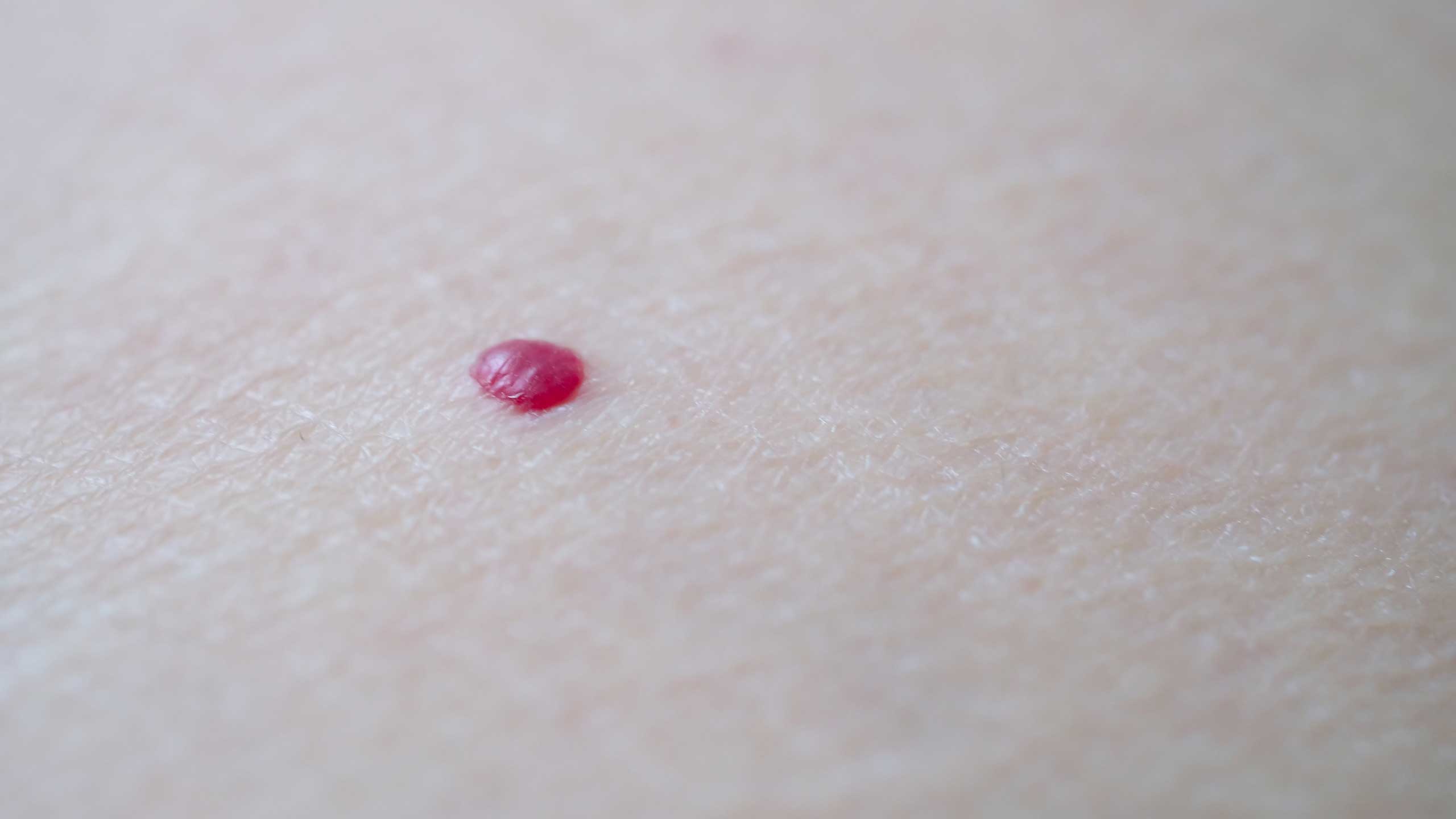
Most of the time, these red spots are a benign vascular lesion called a cherry angioma. The name comes from their round, raised, cherry-red appearance, and once you understand what they actually are, it becomes easy to see why they form and why laser treatment works so well on them.
Why Do Tiny Blood Vessels Clump Together Into a Red Dot?
Underneath the skin, capillaries (blood vessels so fine they're thinner than a strand of hair) spread out in a fine mesh. Normally these vessels stay spread apart, carrying blood evenly across the tissue.
In certain spots, though, several of these thin vessels start to crowd together into one small cluster. When vessels bunch up like this, blood pools inside them, and because the cluster sits close to the skin's surface, it shows through as a red dot. It's a bit like gathering several balloons in one place, the spot where they cluster looks noticeably rounder and more colorful than the rest.
Medically, this kind of vessel cluster is called an angioma. A cherry angioma is specifically a small one sitting close to the surface, which is why it appears as a distinct red dot that doesn't fade when pressed.
Why Does the Number Keep Growing With Age?
Many people start noticing more of these spots from their thirties onward. The reason traces back to how blood vessels themselves change over time.
Vessel walls lose elasticity and grow weaker with age. Once the walls weaken, vessels struggle to hold their original shape, making them more likely to wind through tight spaces or drift close to neighboring vessels. Over time, this naturally leads more small vessels to end up clustered together.
There are also reports that aging shifts the activity of a signaling protein that tells vessels to grow, called vascular endothelial growth factor (VEGF). Think of VEGF as a kind of traffic light for vessel cells, giving the go-ahead to keep growing. When this signal becomes more active in a particular area, it can push new small vessels to form there or encourage existing ones to bunch together. That's why it's so common to see a new spot appear on the torso or arms every so often through your forties and fifties.
Do Genetics and Hormones Play a Role?
If close relatives tend to have a lot of these spots, you're more likely to develop them too. The strength of vessel walls and how prone vessels are to growth both carry a genetic component to some degree.
Hormone shifts are known to matter as well. New cherry angiomas appearing, or existing ones deepening in color, has been reported during pregnancy and other periods of major hormonal change. This is thought to happen because estrogen relaxes blood vessels and makes vessel walls more reactive. That said, there's no evidence linking these spots to cancer or any other systemic disease, so most cases are simply considered a natural part of aging.
How Do You Tell Them Apart From Other Red Marks?
Not every red spot is the same. Some look alike on the surface but have completely different causes, so it helps to know a few distinguishing features.
- Cherry angioma: A distinct red or purplish dome shape that doesn't lose its color under pressure and doesn't fade on its own over time. The vessels are already tightly clustered, so the pooled blood inside can't easily drain out even when pressed.
- Spider angioma (a central vessel with fine capillaries radiating outward like a spider's web): Pressing the center makes the surrounding redness fade too, then it fills back in once you release. This happens because a single central vessel branches into several smaller ones, so pressure briefly cuts off blood flow through those branches.
- Petechiae: Here, blood has leaked out of the vessels into the surrounding skin tissue rather than staying contained inside a vessel, so pressing doesn't change the color at all, and the mark fades over 7 to 14 days, much like a bruise.
Because of these differences, if a red spot suddenly grows larger, becomes asymmetrical, or bleeds easily, it's worth getting it checked, since it could be a different kind of lesion.
How Does Vascular Laser Actually Clear the Spot?
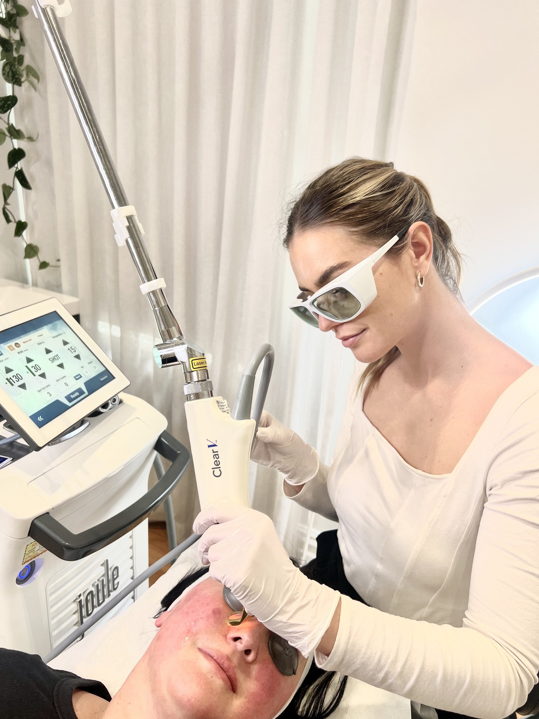
The most common way to remove a cherry angioma is vascular laser treatment, typically a pulsed dye laser (PDL) or an Nd:YAG laser.
These lasers work on a principle called selective photothermolysis. It sounds technical, but the idea is simple. Laser light gets absorbed especially well by substances of a particular color, and hemoglobin (the pigment that makes blood red) happens to absorb this light strongly. It's the same reason a black shirt heats up faster than a white one in the sun.
When the laser hits the vessel, hemoglobin absorbs the light energy and instantly generates heat. That heat transfers to the vessel wall, causing the vessel to coagulate and seal shut. Once sealed, the vessel can no longer carry blood, and the body gradually breaks down and clears away the leftover tissue. The red spot fades bit by bit until it disappears.
The key is that this heat stays concentrated in the vessel itself. Surrounding normal skin cells contain no hemoglobin, so they barely absorb any heat from the same light. That's why only the targeted vessel gets cleared while the surrounding skin stays largely untouched. Reports suggest that smaller lesions closer to the surface tend to respond well after just one or two sessions.
What Does Recovery and Aftercare Look Like?
Right after treatment, the area may look red or slightly swollen. This is a normal reaction as the body absorbs the coagulated vessel tissue.
A small scab sometimes forms at the site, which happens as the surface tissue temporarily breaks down and then heals. Picking at this scab can lead to scarring or discoloration, so it's best to let it fall off on its own.
The treated area can also become more sensitive to sun exposure, so wearing sunscreen for 1 to 2 weeks after treatment is known to help. Avoiding hot compresses and heavy friction on the spot for the same 1 to 2 weeks, until the scab falls off on its own, also helps the healing process along.
Can a Cherry Angioma Come Back After Treatment?
It's rare for the exact same vessel to come back to life after laser treatment, since once it's coagulated and sealed, the body absorbs it completely.
That said, cherry angiomas aren't a condition limited to one spot. They reflect a broader pattern of vessels aging in similar ways across different parts of the body. So even after clearing an existing spot, a new one can appear elsewhere over time. That's not a sign the treatment failed, it simply reflects the ongoing, natural process of vessels aging.
When Should You See a Doctor?
Most cherry angiomas are harmless benign lesions, so there's no medical need to remove them. Still, it's worth getting checked if you notice any of the following.
- A sudden sharp increase in the number of spots over a short period: This can, rarely, be related to other systemic changes worth ruling out.
- A spot that becomes asymmetrical, changes shape, or develops blurred borders: This can signal a different kind of skin lesion that needs to be distinguished from a simple angioma.
- Frequent bleeding or pain from friction against a collar or belt: In spots where irritation keeps recurring, wounds may not heal cleanly, so removing the spot ahead of time is the safer option.
Outside of these situations, removing a cherry angioma for cosmetic reasons with vascular laser is a perfectly reasonable option to consider.
References
Korean Dermatological Association, Ministry of Food and Drug Safety (MFDS), American Academy of Dermatology (AAD), and U.S. Food and Drug Administration (FDA).
Was this helpful?
About this article
Written by a practising aesthetic physician and intended for general education — not a substitute for individual medical advice.
Read next
How Vbeam's Vascular Laser Targets Redness and Broken Capillaries: The Science, Sessions, and Downtime
Vbeam is a vascular laser that uses a specific wavelength tuned to hemoglobin, the pigment in blood, to coagulate dilated capillaries without harming the surrounding skin. This column breaks down why that wavelength zeroes in on blood vessels, along with the bruising, downtime, and sessions it takes to see results.
By Dr. Lee
Those Little Red Spots Called Cherry Angiomas: Why They Appear, and How Vascular Laser or Electrocautery Removes Them Without Scarring
What are those bright red spots called cherry angiomas that show up on the chest or back? A clear, research-backed guide to why they increase with age, how to tell them apart from moles, warts, and purpura, whether pulsed dye, KTP, or long-pulsed Nd:YAG laser or electrocautery works best for removal, how to minimize scarring and dark marks, and when it's time to see a doctor.
By Dr. Kim

Flat Warts, Milia, and Skin Tags: How to Tell Them Apart, the Right Treatment, and Why Removing Them Yourself Is Risky
The small bumps that show up on the face can look alike, but flat warts, milia, and skin tags have different causes and different treatments. Here is how to tell the three apart at a glance, the right CO2 and Er:YAG laser or electrocautery approach for each, how common they are, how they tend to recur, and real evidence on why removing them at home is risky.
By Dr. Lee