Those Little Red Spots Called Cherry Angiomas: Why They Appear, and How Vascular Laser or Electrocautery Removes Them Without Scarring
By Dr. Kim10 min read
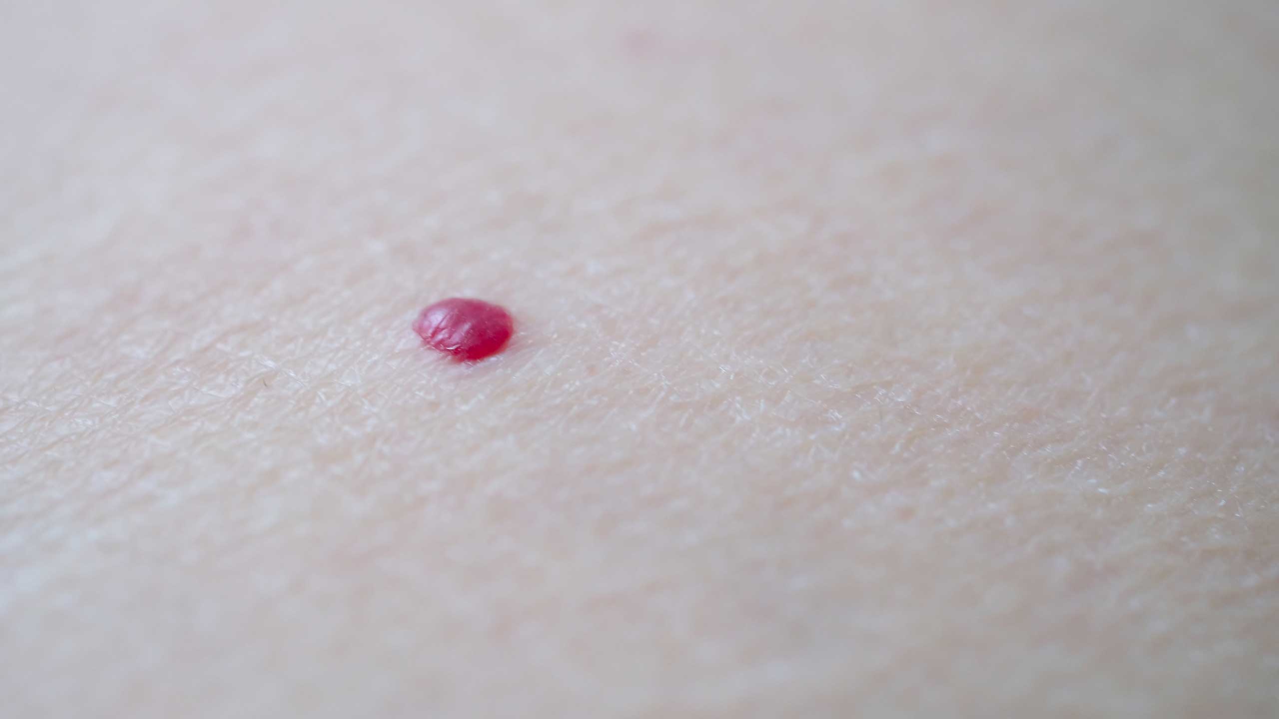
Tiny bright red dots, no bigger than a pinhead, sometimes start showing up on the neck, chest, or arms. They're raised, vivid red, and don't hurt or itch, so many people just leave them alone. But once they start multiplying, or catching on clothing, it's natural to wonder what they actually are. Often, these red spots turn out to be cherry angiomas.
Cherry angiomas, sometimes called senile angiomas, are benign vascular lesions that form when tiny capillaries under the skin cluster together. The good news is that they're completely harmless, and lasers that target blood vessels can usually clear them up nicely. That said, they're easy to mistake for moles, warts, or purpura, and on rare occasions a dangerous lesion can disguise itself as a simple red dot, so it helps to understand what's really going on before deciding how to handle one. Here's what the research actually shows: what a cherry angioma is and why it forms, how to tell it apart from other spots, which lasers work best for removal and aftercare, and when it's time to see a doctor.
So What Exactly Is a Cherry Angioma?
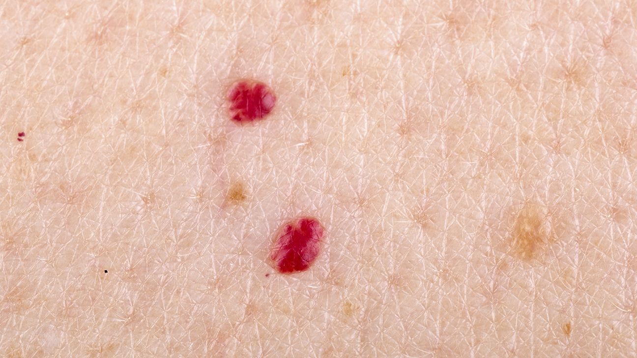
A cherry angioma is a benign lesion that forms when capillaries bunch together in one spot and swell upward. True to its name, it's a bright, cherry red color, with a smooth, slightly domed surface. Most measure just 1 to 5mm across, and they rarely hurt or itch. Because they're essentially a small cluster of blood vessels, though, scratching one with a fingernail or catching it hard on clothing can make it bleed. Cherry angiomas are actually the most common vascular lesion in adults, and they typically start appearing one by one sometime after your thirties.
What you're actually looking at is blood vessels, not pigment, and that's the key difference from a brown or black mole. A mole forms from clustered melanin, the pigment that gives skin its color, while a cherry angioma gets its red glow from blood showing through a cluster of vessels. And here's the reassuring part: it's benign. It won't turn cancerous no matter how long it stays, and it poses no threat to your health.
So a cherry angioma isn't something you need to remove for medical reasons. It really comes down to how cleanly it can be cleared up if it bothers you cosmetically. Still, since its appearance can overlap with other red lesions, the first step is always figuring out exactly what you're dealing with.
How Common Are They, and Why Do They Increase With Age?
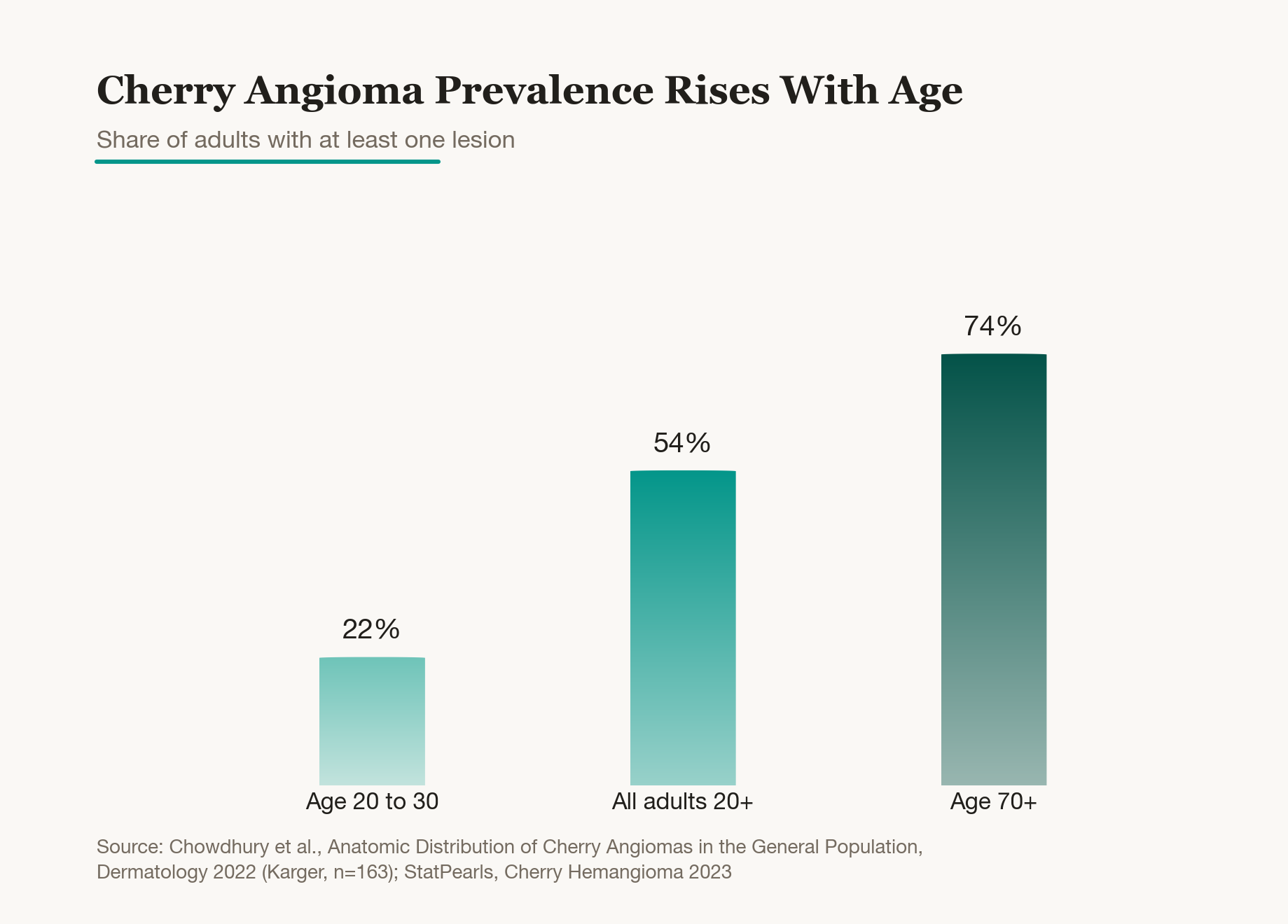
Cherry angiomas are extremely common. In one population survey, 54%, more than half of adults over twenty, had at least one red spot. And the rate rises clearly with age: around 22% in people in their twenties, but well over 70% once people pass seventy.
The number of spots grows with age too. The same survey found that, on average, the count rises by about 48% for every additional decade of life. So the one or two spots you might have had in your twenties often multiply into a scattering across your back and chest by middle age. By the time most people reach their seventies, having several red spots of various sizes is simply the norm.
The exact cause still isn't fully understood. What is clear is that these spots naturally increase with age, and recent research has found mutations in GNAQ and GNA11, genes involved in blood vessel growth, in some lesions. Put simply, it looks like a local switch for building blood vessels gets flipped on, causing capillaries to cluster in one spot. So it's reasonable to think of the occasional new red spot as just another ordinary sign of getting older.
How Do You Tell It Apart From a Mole, a Wart, or Purpura?
It's easy to confuse a red spot with any other kind of raised mark. Since each type differs in color and behavior, correctly identifying it is what determines the right way to treat it.
Color and shape are the biggest clues. A cherry angioma is bright red, raised into a small dome, and stays put for a long time rather than fading after a few days. A mole, by contrast, is brown or black because it's made of melanin, and a wart, caused by an HPV, human papillomavirus, infection, has a rough, bumpy surface and can spread to nearby skin if you scratch it.
Purpura is a bit trickier to tell apart. It happens when a blood vessel ruptures and blood leaks under the skin, spreading out as a flat, red mark much like a bruise. Over time, though, purpura shifts from red to purple to yellow before fading away on its own within one to two weeks. A cherry angioma, on the other hand, doesn't change color or disappear by itself. It just stays there, raised. So if you're looking at a bright red dome that stays firm under pressure and doesn't fade over time, it's likely a cherry angioma. When it's still hard to tell just by looking, a closer exam with magnification, or a biopsy if needed, can settle it.
| Feature | Cherry Angioma | Mole | Wart | Purpura |
|---|---|---|---|---|
| Color | Bright red | Brown, black | Skin tone, brown | Red, changes color |
| Shape | Raised dome | Flat or raised | Rough, bumpy | Flat mark |
| Cause | Capillary clustering | Melanin pigment | HPV infection | Vessel bleeding |
| Course | Persists | Persists | Spreads | Fades in 1-2 weeks |
Where on the Body Do They Show Up, and Why Do They Multiply?
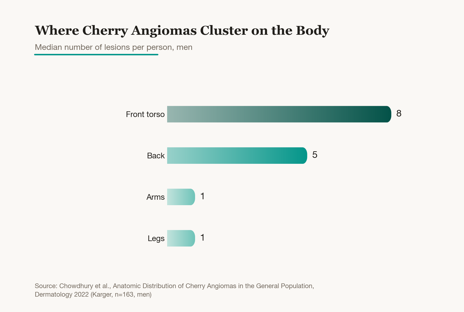
Cherry angiomas can technically appear anywhere on the body, but they do have favorite spots, and the torso is the clear front-runner. In one survey of men, the median count was 8 on the front torso (chest and abdomen), 5 on the back, and only around 1 each on the arms and legs. They rarely show up on the face or neck at all. That's why many people suddenly discover several red spots on their back or chest one day, spots that had simply been hidden under clothing all along. And because the torso isn't somewhere you check often, it's common not to notice until quite a few have already appeared.
A handful of factors influence how many spots you end up with. Age, as mentioned, is the biggest one, but pregnancy and hormonal shifts can also trigger new ones. There are also reports of several spots appearing at once during immunosuppressive treatment or other major changes in the body.
None of this means you should worry just because you have a lot of red spots. Most of the time, this is simply an ordinary part of aging. That said, if a bunch of new ones appear suddenly over a short span, it's less a red flag on its own and more a good prompt to check whether anything else in your body has changed.
How Are They Removed, and How Do You Minimize Scarring and Dark Marks?
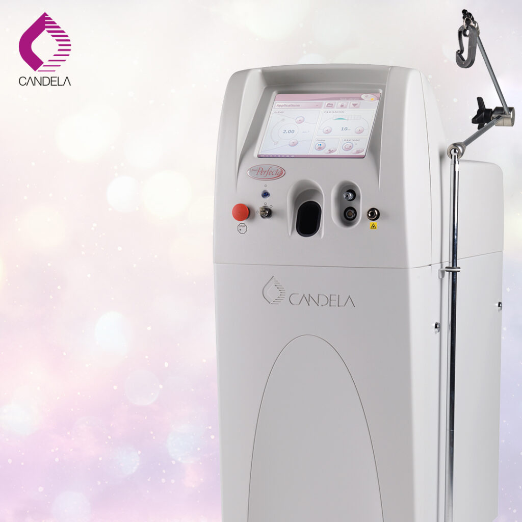
Since a cherry angioma is essentially a bundle of blood vessels, lasers that selectively target vessels are the go-to option. The principle is called selective photothermolysis, a fancy way of saying the laser heats only what absorbs its wavelength. Hemoglobin, the pigment in blood, happens to absorb certain wavelengths of light very well, so when that light hits the lesion, only the blood vessels heat up instantly and seal shut, while the surrounding skin stays largely untouched.
The main options are the pulsed dye laser, the KTP laser at 532nm, and the long-pulsed Nd:YAG laser. Pulsed dye and KTP respond well to shallow red vessels and gently clear small spots. Long-pulsed Nd:YAG has a longer wavelength that reaches deeper, and one comparison study found it needed fewer sessions to clear the lesion. That extra power comes with a trade-off, though: a higher chance of redness or scarring. So KTP or pulsed dye tends to be the safer choice for darker skin tones or more delicate areas, while Nd:YAG really shines on thicker, darker lesions. Electrocautery, which uses fine electrical heat to seal off the vessel, works well for larger lesions since it clears and stops any bleeding in a single step.
The thing to watch most closely afterward is post-inflammatory hyperpigmentation, or dark marks left behind by irritation. Asian skin tends to develop brown marks fairly easily when irritated, so keeping the laser intensity moderate is the first priority. A small scab forms after treatment, and picking at it will leave a mark, so let it fall off on its own and keep up sun protection in the meantime. A spot that's been properly treated won't come back in the same place, though depending on your skin's tendencies, new ones can still pop up elsewhere. If that happens, a light touch-up session handles it.
| Method | Best For | Notes |
|---|---|---|
| Pulsed dye, KTP | Small, shallow spots | Gentle, less pain |
| Long-pulsed Nd:YAG | Thick, dark lesions | Fewer sessions, scar risk |
| Electrocautery | Larger lesions | Stops bleeding at once |

When Should You See a Doctor, and What Signs Matter?
Red spots stay harmless and unchanged for most people's whole lives, and having a lot of them is completely normal. In one survey, 66% of adults had more than 10, 27% had more than 30, and 16% even had more than 50. So the sheer number of spots on your body isn't, by itself, a reason for concern.
A few signs are worth paying attention to, though: a sudden cluster of new spots appearing over a short time, one spot growing unusually fast, or repeated bleeding or scabbing at the same site. An asymmetric, distorted shape or a mottled mix of colors also deserves a look. In very rare cases, a dangerous lesion like amelanotic melanoma, a colorless form of skin cancer, can mimic the look of a simple red spot.
In short, a spot that's changed recently matters far more than one that's been sitting there unchanged for years. If you notice any of these signs, or if new spots are appearing quickly, it's worth getting a checkup before booking a cosmetic removal. There's no need to panic, but there's a real difference between removing something after confirming what it is and removing it without knowing. For the vast majority of people, this is simply an ordinary, harmless change, so it's enough to just keep these warning signs in mind.
Was this helpful?
About this article
Written by a practising aesthetic physician and intended for general education — not a substitute for individual medical advice.
Read next

Milia Removal Around the Eyes: Extraction, CO2 and Erbium YAG Lasers, and Keeping Them from Coming Back
Why those little white bumps around the eyes show up, how milia differ from syringoma, when extraction versus CO2 or erbium YAG laser makes sense, whether you can pick them out at home with a needle, and how likely they are to return, explained simply with real studies.
By Dr. Lee

Flat Wart Removal with CO2 Laser: Why It Spreads, How to Tell It Apart From Milia and Skin Tags, and Managing Recurrence
Flat warts show up as small, flat bumps on the face or back of the hands. Here is why HPV causes them, why scratching makes them spread and become contagious, how to tell them apart from milia and skin tags, how the CO2 laser removes them along with scarring and pigmentation risks, and how to manage recurrence, explained simply with real study data.
By Dr. Lee
How Post-Acne Red Marks Differ from Brown Spots and Scars, and How PDL Laser Treatment Helps
A clear, research-backed guide to why red marks (PIE) appear after acne heals, how they differ from brown pigmentation (PIH) and pitted scars, how long they take to fade naturally, how vascular lasers like Vbeam and Excel V speed up the process, how to manage them at home with sunscreen and calming care, and why treating them early matters before they turn into scars.
By Dr. Kim