How CO2 Laser Mole Removal Works: Recurrence, Scarring, Pigmentation, and the ABCDE Signs of Melanoma
By Dr. Kim11 min read

A lot of people go back and forth on whether to have a mole removed, whether it sits on the face, the neck, or the back of a hand. Makeup does not always cover it well, and when it keeps catching on a razor or a washcloth, it starts to bother you. Once you look into mole removal, CO2 laser is usually the first option that comes up.
Most moles are harmless, benign growths, so once you understand how they work, clearing one up is fairly straightforward. But not every mole is the same. A shallow mole and a deep one call for different removal approaches. And on rare occasions, something as serious as melanoma can look just like an ordinary mole, so there are warning signs worth checking before removing anything without a second thought.
Here is a closer look at what a mole actually is, how it differs from seborrheic keratosis, skin tags, and warts, how CO2 laser removes it, how recurrence and scarring are managed, how deeper moles are treated, and exactly when you should see a doctor, all backed by real research numbers.
What Exactly Is a Mole?
A mole forms when melanocytes, the cells that produce melanin, cluster together in one spot. In medical terms it is called a melanocytic nevus. In plain language, it is simply a cluster of pigment-producing cells settling in one place, and in most cases it poses no threat to your health.
Moles fall into three types depending on how deep the cells sit. When the cells sit shallow, right at the border between the epidermis and dermis, it is called a junctional nevus, and it tends to be flat and fairly dark. When the cells span both the border and the dermis, it is a compound nevus, which sits slightly raised. And when the cells settle deep in the dermis, it is a dermal nevus, which rises as a plump, dome-shaped bump in skin tone or light brown.
Some moles are present from birth, while others appear later with age or years of sun exposure. That is why the number varies from person to person, and it tends to increase as people move into adulthood.
This depth matters for a simple reason. A shallow mole clears up cleanly even with a light pass of the laser, but a mole rooted deep in the dermis can leave cells behind if you only treat the surface, and it is likely to resurface. So the first step before removal is figuring out whether your mole sits shallow or deep.
| Type | Depth | Shape | Color |
|---|---|---|---|
| Junctional nevus | Epidermal border | Flat | Dark brown to black |
| Compound nevus | Border and dermis | Slightly raised | Brown |
| Dermal nevus | Deep in dermis | Raised, dome shaped | Skin tone to light brown |
How Do You Tell a Mole Apart From Seborrheic Keratosis, Skin Tags, and Warts?
Not every brown spot on the face or neck is a mole. Seborrheic keratosis, skin tags, and warts are just as common, and they are easy to mix up, but each has a different cause and a different nature, so figuring out which one you have determines the right way to treat it.
A mole, as described above, is a melanocytic nevus formed by clustered pigment cells. Seborrheic keratosis, on the other hand, is a brown patch caused by sun exposure and aging thickening the epidermis, which makes it a problem with keratin and skin surface cells rather than pigment cells. A skin tag is a soft, skin-colored growth that hangs off the skin, caused by friction and aging loosening the tissue, and it has nothing to do with pigment. A wart comes from HPV, the human papillomavirus, and it has a rough, bumpy surface. Scratching one can spread it to nearby skin, and it can also spread to other people.
The distinction matters because the treatment differs for each. Moles are treated with laser or excision depending on depth, seborrheic keratosis with a light pigment laser, skin tags with laser cauterization, and warts with cryotherapy or cauterization. When the appearance alone is not enough to tell, magnification or, if needed, a biopsy can confirm it. Identifying what you actually have first helps avoid unnecessary procedures.
| Mole | Seborrheic Keratosis | Skin Tag | Wart | |
|---|---|---|---|---|
| Cause | Melanocytic nevus | Sun exposure and aging | Friction and aging | HPV infection |
| Appearance | Flat or raised | Flat brown patch | Soft, hanging stalk | Rough, bumpy papule |
| Contagious | No | No | No | Yes, can spread |
| Treatment | Laser or excision, by depth | Pigment laser | Laser cauterization | Cryotherapy or cauterization |

How Does CO2 Laser Remove a Mole?
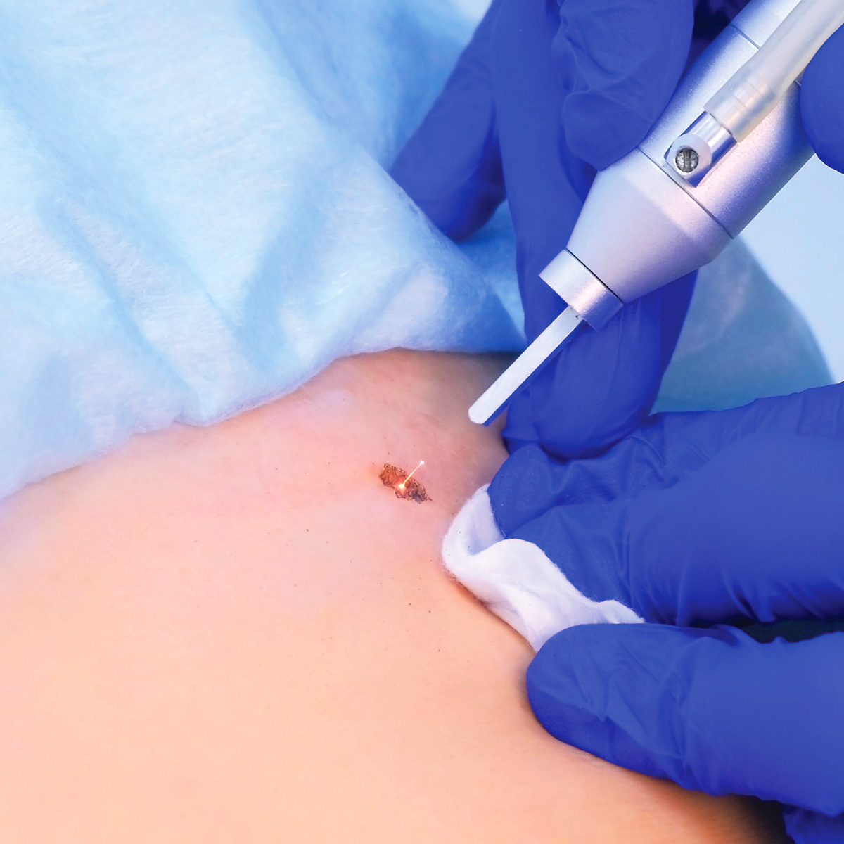
CO2 laser is the standard workhorse for mole removal. It fires light at a 10,600nm wavelength, which water absorbs extremely well. Since most of our skin tissue is made up of water, the moment the laser energy hits, the moisture in that spot boils instantly and the tissue evaporates. This is called ablation. In simple terms, it burns off the thin layer where the mole sits.
The advantage of this ablation process is that it can shave away extremely thin layers at a time. Each pass of the laser evaporates a little more of the surface, so the treatment can be broken into several light passes matched to how deep the mole runs. A shallow junctional nevus usually clears in just one or two passes, while a raised compound nevus needs a bit more shaving. On top of that, the heat seals off tiny blood vessels as it works, so there is very little bleeding and the finish stays clean.
Still, depth control is what makes or breaks the result. Burn too deep in one pass and the mole is gone for sure, but scarring becomes more likely. Go too shallow, and cells are left behind to resurface later. That is why the practitioner's judgment, matching intensity to the type and depth of the mole, matters so much. Right after treatment, a small scab forms over the spot, and as that scab falls away, the mole clears in a natural process. In the end, the result depends on the device's mechanism, the practitioner's skill, and proper aftercare all coming together.

Does a Mole Come Back After Removal?
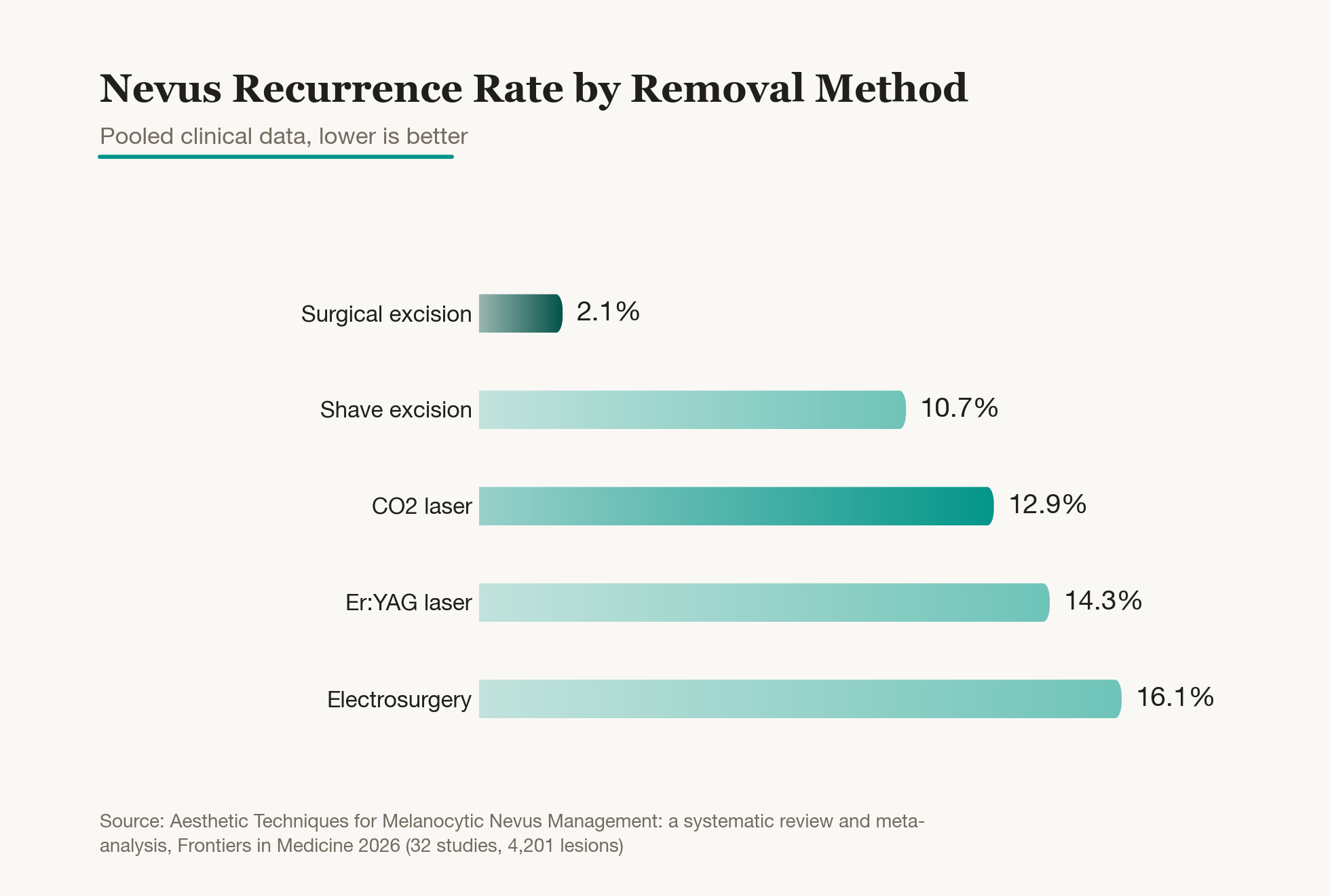
Recurrence and scarring are the two biggest worries after mole removal. To be honest, CO2 laser is not a method with zero chance of recurrence. A pooled analysis of multiple studies found that moles removed with CO2 laser came back at a rate of about 13%. Surgical excision with a scalpel, by comparison, had the lowest recurrence at about 2%, with laser running higher.
That does not make laser a bad choice. In the same analysis, scarring occurred in 21% of surgical excision cases, the highest of the two, compared with just 5% for CO2 laser. In other words, laser trades a slightly higher recurrence risk for a much lower chance of scarring, while excision trades a higher chance of scarring for a near-certain end to recurrence. So the right choice depends on how deep and where the mole sits, and whether that spot is somewhere a scar would be noticeable.
Pigmentation shows up almost as often as recurrence. Asian skin tends to mark easily with brown discoloration after irritation, and pigmentation after CO2 laser is reported in about 9% of cases. Most of these marks fade with time, but sun exposure makes them darker and slower to clear. That is why not picking at the scab and staying consistent with sun protection during recovery makes up half the outcome. If cells remain after a shallow pass and the mole resurfaces, it can be touched up again once healing is confirmed.
How Are Deep or Large Moles Removed?
Not every mole is treated with laser alone. A plump mole rooted deep in the dermis, or one that is simply large, tends to leave cells behind and resurface if only the surface is lasered away. For moles like these, excision that removes everything down to the root from the start gives a cleaner result.
Small, round moles are handled with punch excision. A round blade cuts the mole out down to its base, and one or two stitches close the spot, so even a deep mole is cleared completely in a single pass without leaving cells behind. It is done under local anesthesia, so the discomfort is minimal, and for a small mole the procedure itself takes only a few minutes. If the mole is larger or elongated, an elliptical excision is used instead, cutting a longer shape and closing it with sutures. The removed tissue can also be sent for pathology, which has the added benefit of ruling out any abnormal cells.
Of course, excision means stitches come out a few days later and there is a healing period, and it leaves a fine scar. Even so, because everything is cleared down to the root, recurrence is much lower. So the general rule is laser for shallow moles, which avoids scarring, and excision for deep or large moles, which avoids recurrence. Neither is unconditionally better. It comes down to matching the method to the mole's depth, size, and location. In particular, if a longstanding mole suddenly grows larger or changes shape, it is safer to consider excision first, since it allows tissue to be checked, before removing it for cosmetic reasons alone.
When Should You See a Doctor, and How Do You Recognize Melanoma?
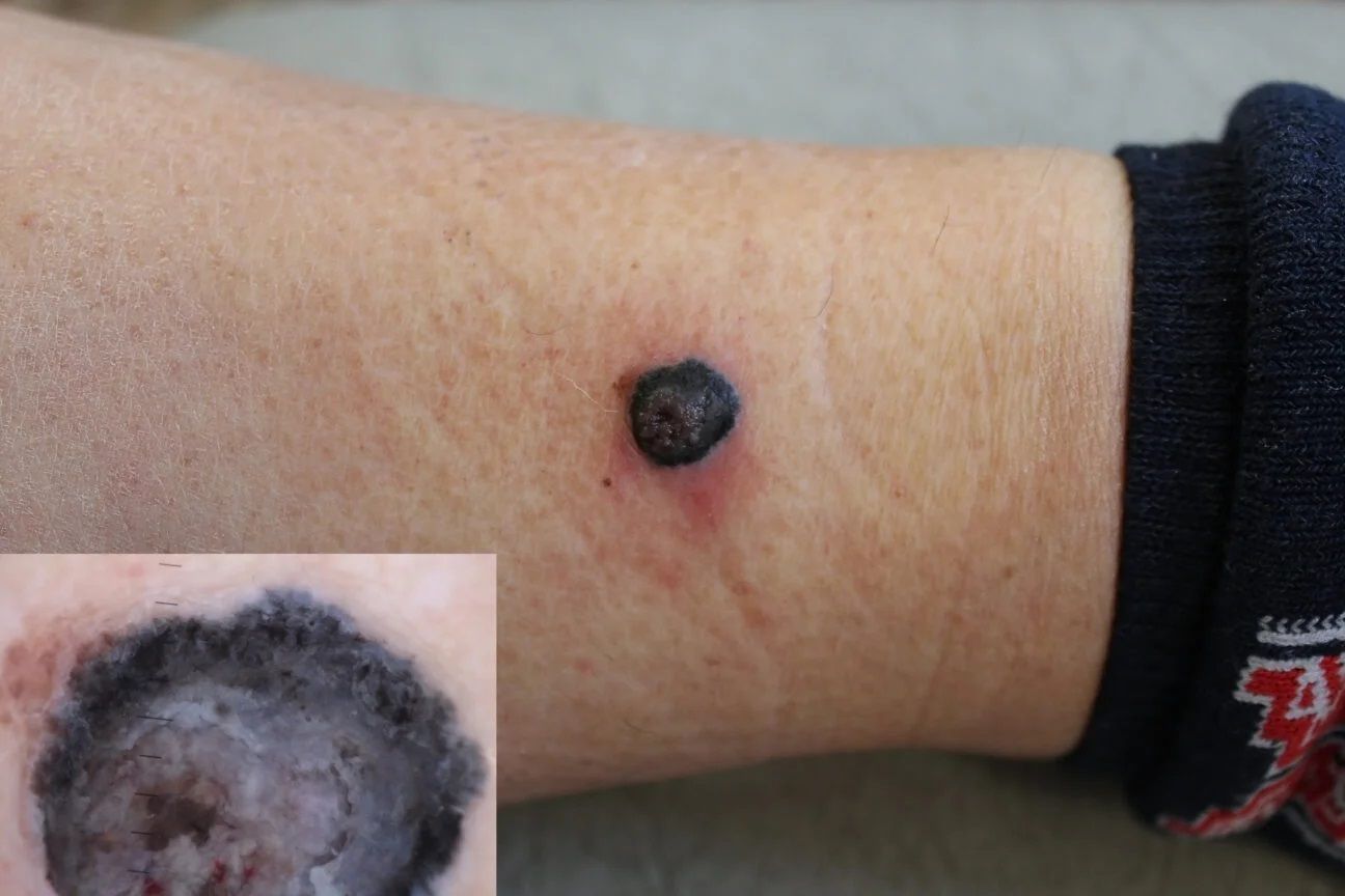
Nearly every mole stays a quiet, benign growth for life. But on rare occasions, melanoma, a form of skin cancer, can look just like an ordinary mole, so there are warning signs worth checking before removal. The ABCDE rule is a simple way to remember them.
A stands for asymmetry, meaning the two halves do not match if you fold the mole in half. B stands for border, meaning the edges are ragged or blurred. C stands for color, meaning brown, black, and red are mixed unevenly within one spot. D stands for diameter, meaning it measures more than 6mm across. And E stands for evolving, meaning a mole that has recently grown, changed color, started itching, or begun to bleed.
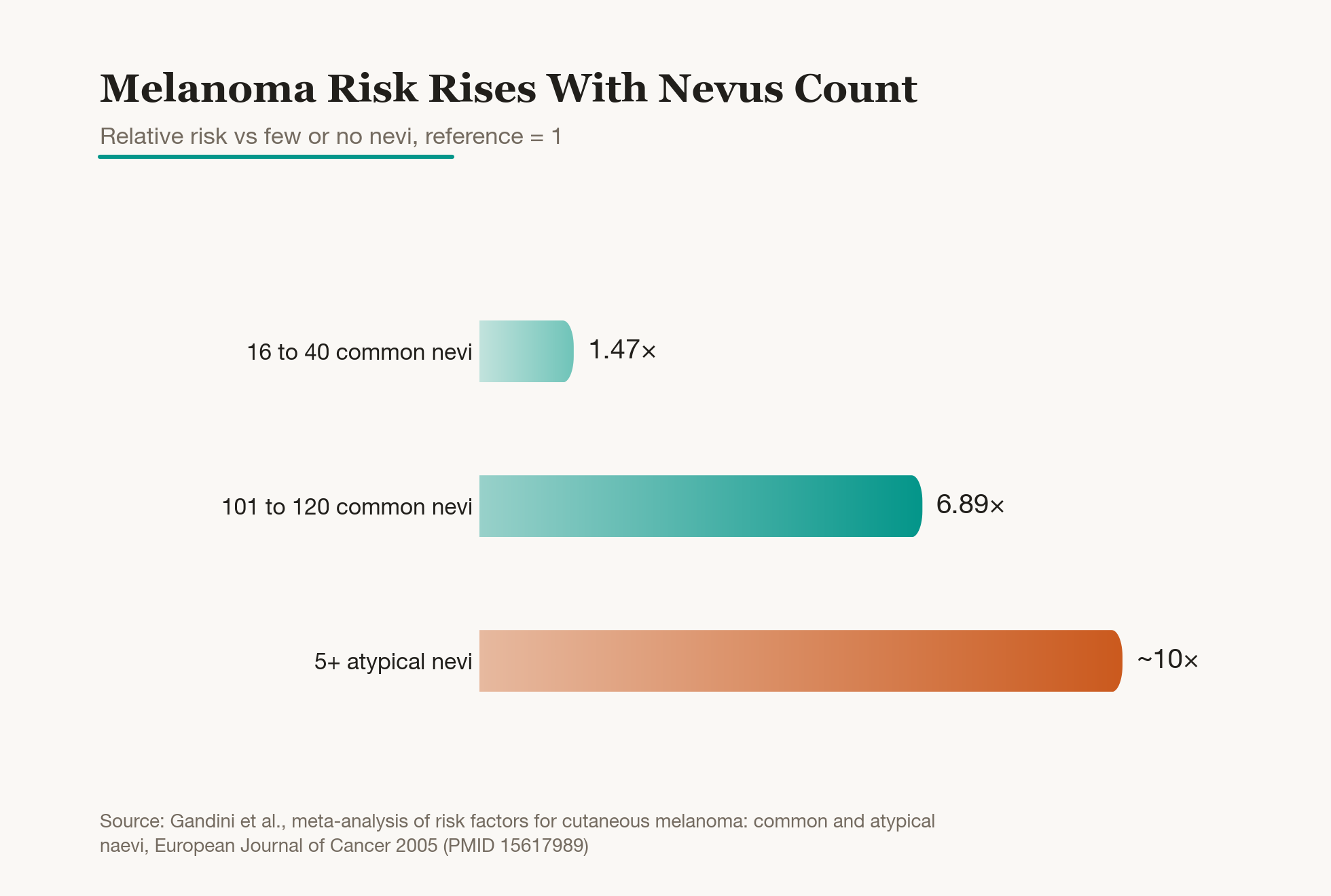
Of these, evolving is the most important. A mole that suddenly changes is far more concerning than one that has stayed the same for years. Having a lot of moles is itself linked to melanoma risk, too. One analysis found that having more than 100 moles raises the risk by about 7 times, and having several irregularly shaped, atypical moles raises it by about 10 times. So if any of these signs show up, or if your mole count starts climbing quickly, it is safer to see a doctor first before removing anything for cosmetic reasons. There is no need to panic, but getting checked before removal and removing one without checking are clearly not the same thing.
| Letter | Meaning | Warning Sign |
|---|---|---|
| A, Asymmetry | Asymmetry | The two halves do not match |
| B, Border | Border | Edges are ragged or blurred |
| C, Color | Color | Multiple colors mixed together |
| D, Diameter | Diameter | Larger than 6mm across |
| E, Evolving | Evolving | Recently grew or changed |
Was this helpful?
About this article
Written by a practising aesthetic physician and intended for general education — not a substitute for individual medical advice.
Read next
Those Little Red Spots Called Cherry Angiomas: Why They Appear, and How Vascular Laser or Electrocautery Removes Them Without Scarring
What are those bright red spots called cherry angiomas that show up on the chest or back? A clear, research-backed guide to why they increase with age, how to tell them apart from moles, warts, and purpura, whether pulsed dye, KTP, or long-pulsed Nd:YAG laser or electrocautery works best for removal, how to minimize scarring and dark marks, and when it's time to see a doctor.
By Dr. Kim

Flat Wart Removal with CO2 Laser: Why It Spreads, How to Tell It Apart From Milia and Skin Tags, and Managing Recurrence
Flat warts show up as small, flat bumps on the face or back of the hands. Here is why HPV causes them, why scratching makes them spread and become contagious, how to tell them apart from milia and skin tags, how the CO2 laser removes them along with scarring and pigmentation risks, and how to manage recurrence, explained simply with real study data.
By Dr. Lee

Those Tiny Bumps Spreading Across Your Face: Why Picking at Flat Warts Only Makes Them Multiply
Flat warts are a viral skin condition, so picking or scratching at them doesn't clear them up, it spreads them further. Here's why that happens, how to tell them apart from moles or acne, and the logic behind removal and preventing recurrence.
By Dr. Kim